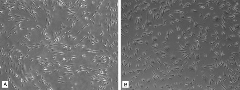
Figure 2.

Cell morphology and attachment. HUVECs exhibited different morpholgy when grown on two different conditions. They exhibited a large and spindle shape, and random organized on six-well TCP (B). Compared to the TCP sample, the cells on ECM were much smaller and elongated, with a regular arrangement following the ECM organization (A). (Original magnification 5×).
