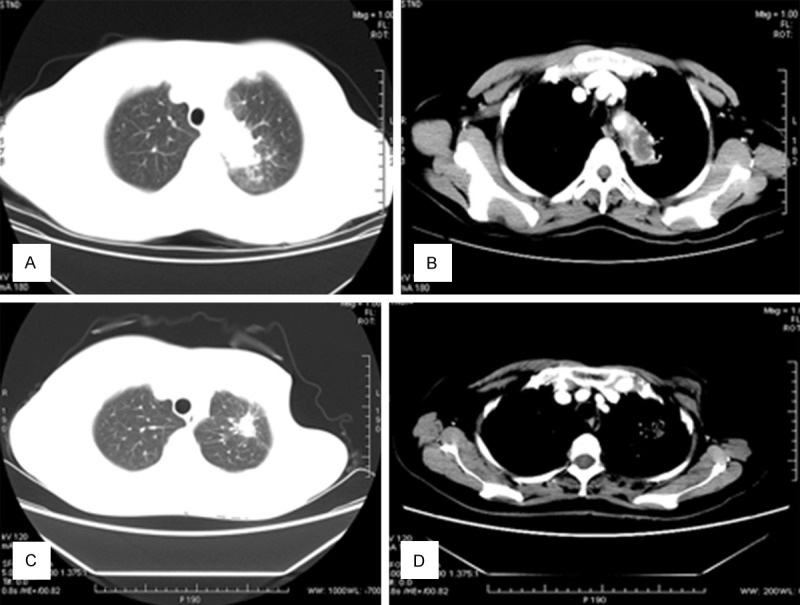
Figure 1.

A, B. A 43-year-old woman with hemoptysis with no obvious cause. Chest CT shows a nodule with lobulation, and the halo sign. Intravenous contrast-enhanced CT reveals that the nodule is heterogeneously enhanced and contains an area of low density (necrosis), measuring 2×2.5 cm. The postoperative pathological examination revealed an Aspergillus infection. C, D. A 56-year old woman was diagnosed with breast cancer and underwent surgery 11 years ago. Thereafter, she had underwent several cycles of chemotherapy. Three years ago, she developed intermittent hemoptysis. CT shows a lesion with high density and an irregular boundary and spiculation in the left upper lung, surrounded by patchy. In the mediastinal window, the lesion is partly subtracted.
