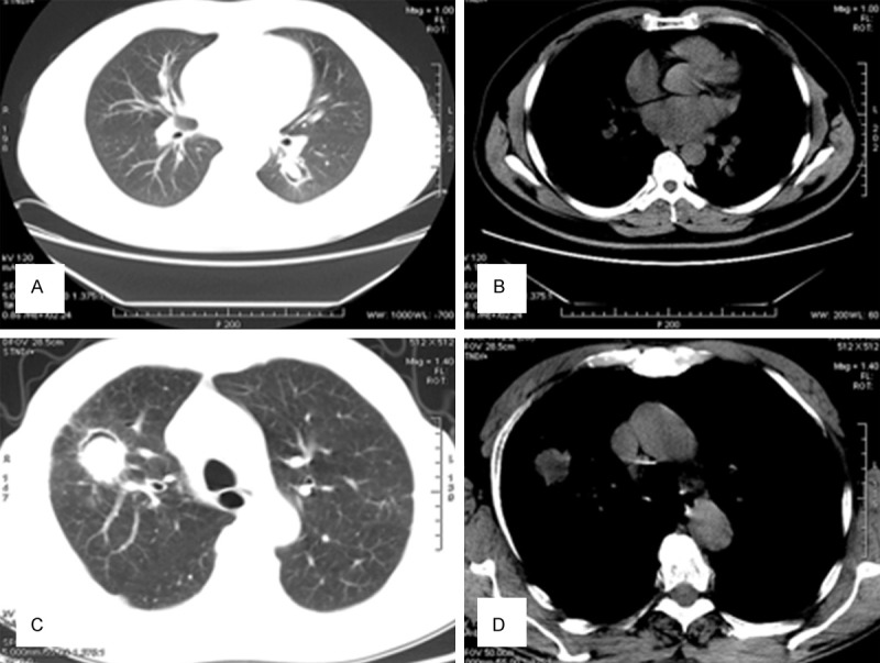
Figure 2.

A, B. A 35-year old woman with a more than 2-year history of hemoptysis. A mass is observed in the right lung. The lesion has a clear boundary and lobulation, and measures 3×2.5 cm. C, D. In the right lung, consolidation is observed with an ill-defined border and uneven density, within which cavity formation can also be observed.
