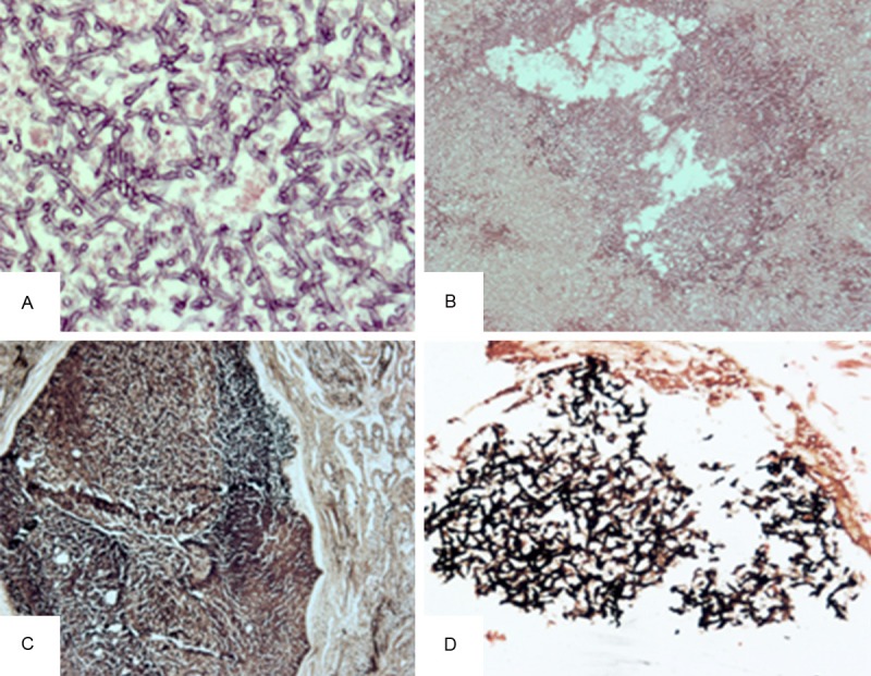
Figure 4.

A, B. Aspergillus hyphae on HE staining. There are numerous Aspergillus hyphae with 45° branching (A: original magnification, ×400; B: original magnification, ×200). C. The lesion is surrounded by fibrous tissue (original magnification, ×100). D. GMS staining shows fungal elements (original magnification, ×200).
