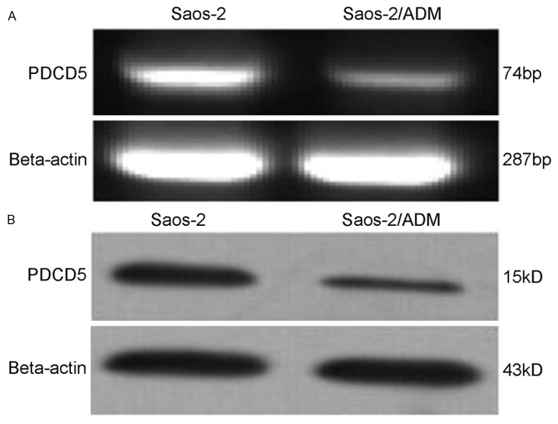
Figure 1.

Expression of PDCD5 in Saos-2 osteosarcoma as detected by the RT-PCR (A) and Western blotting (B). PCR products were electrophoresed on a 2.0% agarose gel, with beta-actin as an internal control. For Western blotting, cell lysates were separated on a 12% SDS polyacrylamide gel, and the blot was probed with a specific monoclonal anti-PDCD5 antibody, with beta-actin as an internal standard to normalize loading protein.
