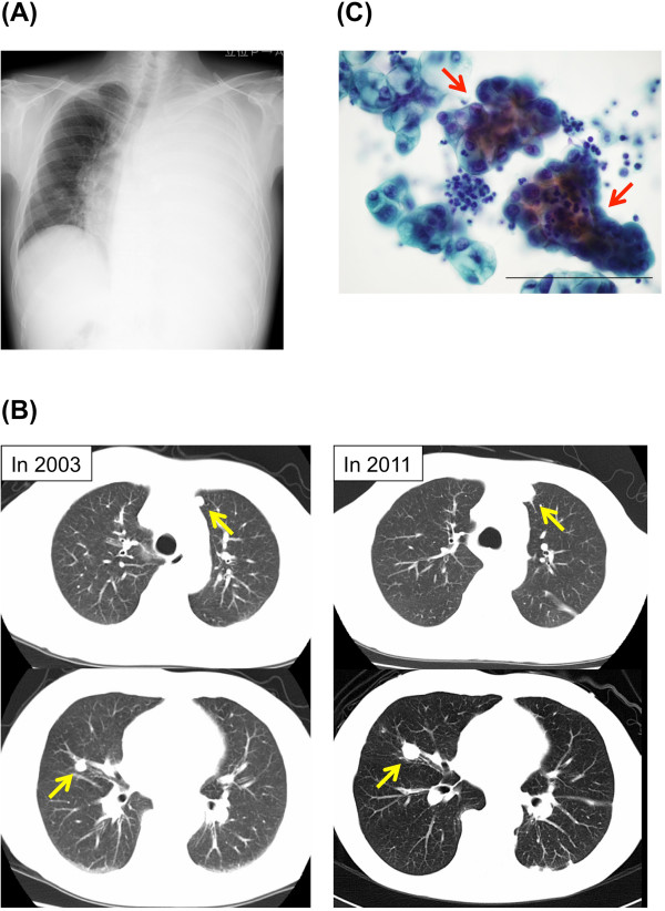
Figure 1.

Clinical findings of the case. (A) Chest X-ray obtained on admission in 2011 shows a massive left-sided pleural effusion. (B) Multiple lung nodules (yellow arrows) are visible on computed tomographic scans obtained in 2003 (left images) and on admission in 2011 (right images, after drainage of pleural effusion). (C) Cytological findings of the pleural effusion. Cell clusters with peripherally located irregular nuclei (red arrows) are shown. Scale bar: 100μm.
