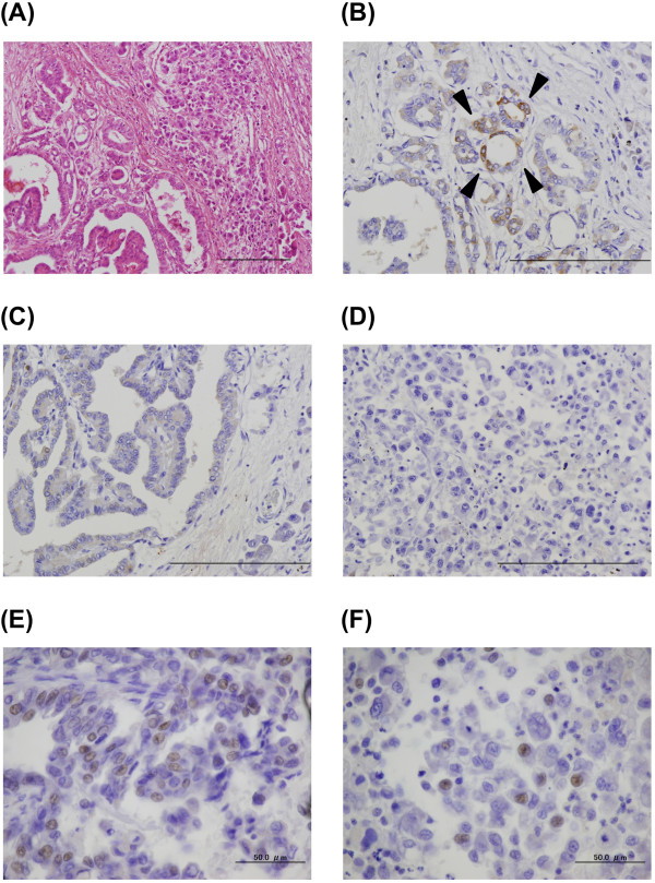
Figure 2.

Microscopic findings of the lung metastatic tumor at autopsy. (A) Hematoxylin and eosin stain. A transitional zone between the differentiated papillary pattern (left side of the image) and the undifferentiated component (right side of the image) is shown. (B, C, D) Immunohistochemical staining for thyroglobulin. Differentiated papillary carcinoma positive (B, arrowheads) and negative (C) for thyroglobulin and an undifferentiated component negative for thyroglobulin (D) are shown. (E, F) Immunohistochemical staining for thyroid transcription factor 1. Differentiated papillary carcinoma positive for thyroid transcription factor 1 (E) and an undifferentiated component positive for thyroid transcription factor 1 (F) are shown. Scale bars: 200μm (A-D), 50μm (E, F).
