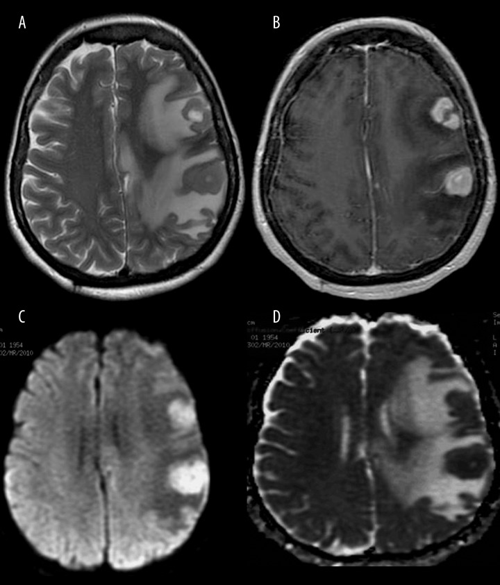
Figure 12.
Primary central nervous system lymphoma. MR images show two homogeneously hypointense tumors on T2-weighted image (A) with strong contrast enhancement on post-contrast T1-weighted image (B). DWI reveals almost homogenous diffusion restriction with high signal on DW image (C) and low signal on the ADC map (D).

