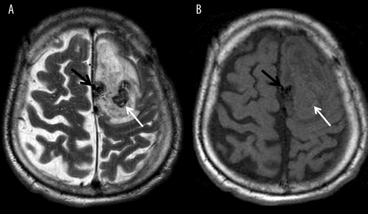
Figure 2.
Intracerebral active bleeding from an arteriovenous malformation located parasagitally (black arrows) within the left hemisphere; (A) T2-weighted and (B) T1-weighted images. Central area of low signal on T2-weighted image (A) is consistent with acute bleeding and deoxyhemoglobin (white arrows) which is surrounded by a large hyperacute hematoma with T2 and T1 signal characteristic of oxyhemoglobin.

