Figure 6.

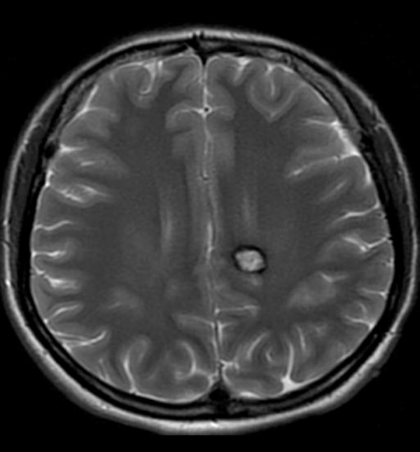
Cavernoma in the left parasagittal location. T2-weighted image shows typical salt and pepper appearance with central high signal and peripheral hypointense rim.

Cavernoma in the left parasagittal location. T2-weighted image shows typical salt and pepper appearance with central high signal and peripheral hypointense rim.