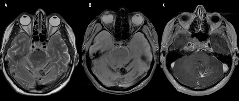
Figure 7.
Cavernoma and developmental venous anomaly within the left cerebellar hemisphere. T2-weighted image (A) shows hypointense oval cavernoma and bands of superficial hemosiderosis due to chronic bleeding which are better depicted on SWI (B). Contrast-enhanced T1-weighted image (C) reveals coexisting developmental venous anomaly.

