
Fig. 5.
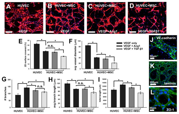
Generation of microvascular networks in the presence of BM-hMSC and different biomolecules. Initially uniformly dispersed HUVECs form perfusable networks by vasculogenesis-like process in VEGF supplemented standard endothelial growth medium with and without BM-hMSCs or with VEGF+Ang-1 and BM-hMSCs (A–D). Microvascular networks under various conditions are quantified E–I). Functionality of microvessel sare confirmed by staining with VE-cadherin (J, green) for adherens junctions, laminin (K, green) for matrix secretion, and ZO-1 (L, green)for tight junctions. Cell nuclei were stained with DAPI (blue). Scale bars = 200μm, unless noted.