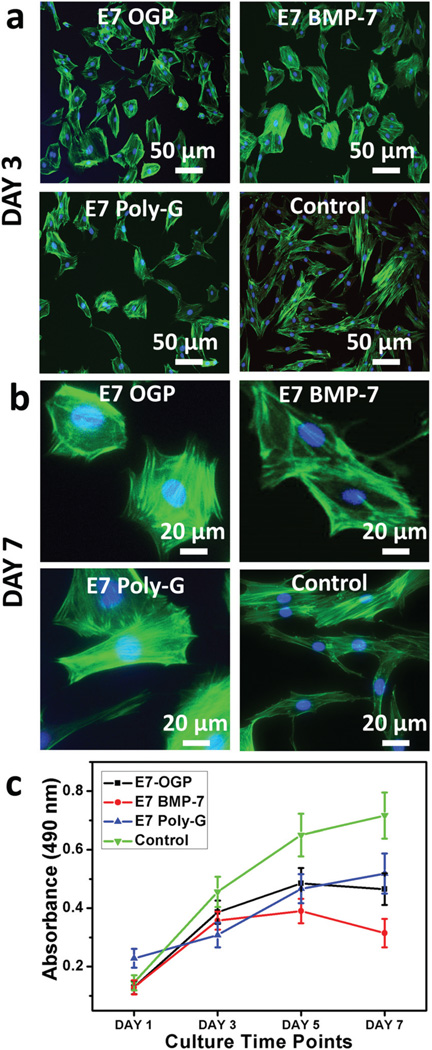
Fig. 5.
Cell adhesion and proliferation on different substrates. Confocal images showing cell adhesion on the different peptide-coated HA surfaces and cell proliferation after cell seeding for three days (a) and seven days (b). All materials exhibited high biocompatibility and strongly supported cell adhesion. Cells presented a significant fibroblast-like morphology in the groups of blank control and E7 Poly-G. The cell morphology changed to the broad shape in the group of E7 OGP and E7 BMP-7. Cell nuclei were stained by DAPI (blue) and F-actin was stained by FITC-labelled phalloidin (green). Cell proliferation over time is shown (c) on the different peptide-modified discs.

