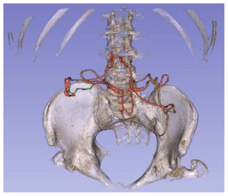
Fig. 4.

3D segmentation of the marginal artery with pelvis and spine for reference. The artery was tracked following the transverse and descending colon. The portion shown communicates between the SMA and IMA. Ground truth is labeled in green, and SMC detection is labeled in red. Detection shown has recall of 94.9% and precision of 58.3%.
