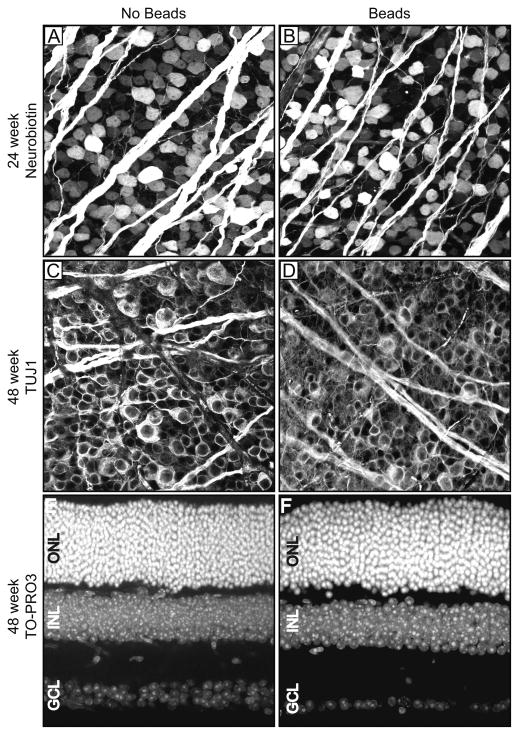
Figure 2. Prolonged IOP elevation results in RGC loss.
Examples of uninjected eyes (“No Beads”; panels A, C, E) and bead-injected eyes (“Beads”; panels B, D, F) are presented side by side at 24 weeks after injection (panels A and B), or 48 weeks after injection (panels C–F). A–D. Retinal flat mounts. E, F. Retinal cross sections. A, B. Neurobiotin-staining. C, D. TUJ1-staining. E, F. TO-PRO3 staining. An extreme example of RGC loss (F) is shown in comparison with a typical control retina (E). The INL and ONL are of equivalent thickness. ONL = outer nuclear layer; INL = inner nuclear layer; GCL = ganglion cell layer.

