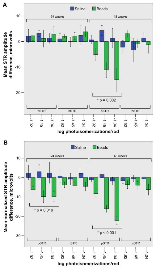
Figure 4. Prolonged IOP elevation results in a decrease of the pSTR amplitude difference.
A and B. The mean pSTR and nSTR amplitude difference (lower brackets) following stimulation at three distinct scotopic light intensities (log photoisomerizations/rod) of animals exposed to either 24 or 48 weeks of IOP elevation (upper brackets) is provided. Saline (blue) and bead (green) injected animals of the same exposure duration and light intensity are presented side by side. A. At 24 weeks of IOP exposure there is no reduction in either the pSTR or the nSTR amplitude difference in bead-injected animals when compared to saline-injected animals. However, at 48 weeks of IOP exposure, the pSTR amplitude difference is diminished whereas the nSTR amplitude difference is not. B. At all light intensities at both time points the normalized pSTR amplitude difference is reduced in bead-injected animals when compared to saline-injected animals. For both panels, an asterisk denotes an ANOVA with repeated measures with the indicated p value. One SEM is shown. Note the difference in the scale of the y axis between the two panels.

