Fig. 6.

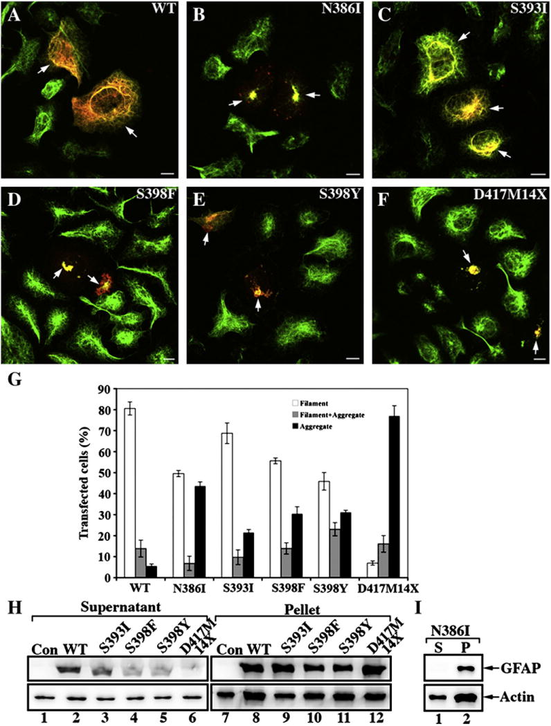
Transient expression of mutant GFAPs leads to aggregate formation in SW13/cl.1 (Vim+) cells. SW13/cl.1 (Vim+) cells transiently transfected with either WT (A) or mutant (B–F) GFAP were processed for double label immunofluorescence microscopy with antibodiesto both GFAP (red channel) and vimentin (green channel). For each transfection, cells on three coverslips were counted and their staining patterns (Filament; Filament + Aggregate; Aggregate) assessed. Quantification of the various patterns observed for transfected SW13/cl.1 (Vim+) cells are shown as mean ± SE and presented as bar charts (G). All the GFAP mutants resulted in a significant increase in GFAP aggregation in transiently transfected cells with the D417M14X deletion mutant predominantly forming protein aggregates. Bar=10 μm. (H,I) SW13/cl.1 (Vim+) cells transfected with the indicated GFAP construct were then extracted using a mild extraction protocol. The resulting supernatant (S) and pellet (P) fractions were analyzed by SDS-PAGE followed by immunoblotting with GA5 (H) and SMI-21 (I) monoclonal anti-GFAP antibodies. Actin was used as a sample loading control. Uncropped images of blots are shown in Supplementary Fig. 2.
