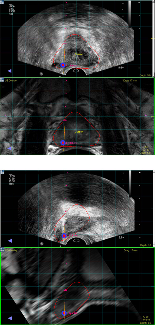
Figure 7.
MRI registered with ultrasound visualized in the axial plane (a) and sagittal plane (b). A biopsy target is located in the left apical mid area of the prostate and visualized in red. Needle trajectory is shown by the red dots, and the orange line maps the biopsy location for archiving and later use.

