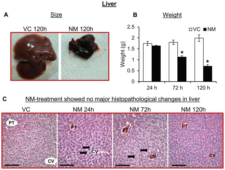
Figure 6. Topical application of NM on to the dorsal skin of SKH-1 hairless mice causes decrease in size and weight of liver.
Dorsal skin of mice was exposed topically to either NM (3.2 mg) in 200 μL acetone or 200 μL of acetone alone. 24, 72 and 120 h post-NM exposure, liver was collected, processed, sectioned and subjected to H&E staining as detailed under ‘Materials and Methods’ section. NM-induced changes in the size and weight of the liver were recorded (A and B) and liver histopathology was analyzed (representative pictures, C). VC, vehicle control (acetone); NM, nitrogen mustard-exposed mice; PT, portal triad; CV, central vein; black arrows, dilated sinusoids; red scale bar, 1 cm; black scale bar (histology images), 100 μm. Data represent mean ± SEM of three-eight animals in each treatment group. *, p < 0.05.

