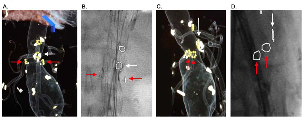
Figure 1. 3D Overlay Fusion and Overlay Imaging.
This image demonstrates the method of overlay mark production and utilization. Panel A demonstrates the marks placed around the origins of the SMA (white arrow) and renal arteries (red arrows). Panel B demonstrates intraoperative overlay of these marks on live fluoroscopy. The radiopaque markers on the graft can be seen to closely approximate these overlay marks during deployment. Panel C and D demonstrate the same patient in a lateral projection. Note that a perfectly orthogonal image of the vessel origin can be obtained by aligning the origin marker such that it appears as a line on the overlay, and this can be aligned before initiating fluoroscopy, minimizing radiation during adjustment of the c-arm.

