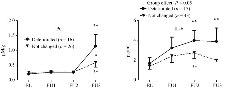
Figure 1. Comparison of protein carbonylation and IL-6 levels during follow-up in patients whose LV diastolic function remained unchanged or deteriorated.
*P < 0.05, **P < 0.01. The table includes the results of only those patients who had at least three measurements of the investigated parameters during the follow-up. BL: baseline; IL-6: interleukin-6; FU: follow-up; LV: left ventricular; PC: protein carbonylation.

