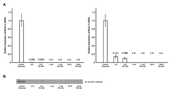
Fig. 2.
A) qRT-PCR. The expression level of Mmp 1a (left) and Mmp 1b (right) after 24 hr exposure to CSE was analyzed by qRT-PCR. The expression level was normalized by internal control and shown its expression relative to control sample (mouse placenta as 1). There was no induction of Mmp 1a nor Mmp 1b after 12 and 72 hr exposure as well as serum starved condition (data not shown). B) Western blotting. Expression of MMP1 after 24 hr exposure was confirmed by western blotting. The serum free culture media from cells were collected and applied to the lanes. Tissue extract from mouse placenta was used as positive control. There was no induction of Mmp 1a nor Mmp 1b after 72 hr exposure (data not shown).

