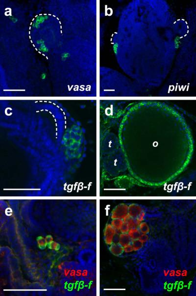
Figure 5. Expression patterns of markers for germline and germline-associated cells in B. schlosseri.

(a) vasa is expressed by clusters of germ cells associated with the secondary bud (dashed line) and just posterior to the secondary bud at stage B2. (b) At stage B1, piwi-positive germ cells are present posterior to the secondary buds (dashed lines). (c-d) tgfβ-f expression is detected in clusters of primitive follicle cells posterior to the secondary bud (dashed lines) at stage A2 in non-fertile juveniles (c) and in follicle cells surrounding the maturing oocytes (o) and testes (t) in the primary buds of fertile animals (d). (e-f) Two-color FISH can be used to simultaneously visualize vasa expression (red) and tgfβ-f expression (green) in non-fertile (e) and fertile (f) animals. Scale bars indicate 50 μm.
