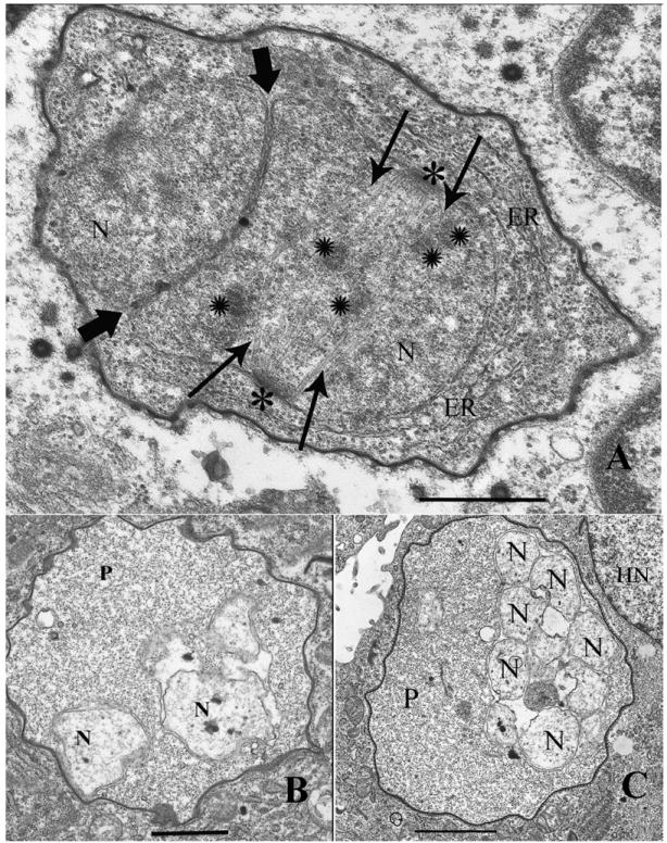
Fig. 2.

Electron microscopic observations of A. algerae development in control RK13 cultures and in albendazole treated infected cells. A. Infected (Control) RK13 cell with A. algerae developing in its cytoplasm. The typical proliferative stage of the parasite contains endoplasmic reticulum (ER), nuclei (N) in a diplokaryotic arrangement with tightly abutted nuclear membranes (bold arrowheads). The diplokaryon is undergoing karyokinesis, which is evident due to the presence of nuclear spindle plaques (*), spindle microtubules (arrows), and chromosomes (✺). Magnification bar = 600nm. B. Albendazole treated RK13 cell infected with an A. algerae proliferative (P) stage developing in its cytoplasm. The parasite has a homogeneous granular cytoplasm, lacking ER or any other defining structure. The nuclei (N) are clearly degenerate having separated; they are no longer in its characteristic diplokaryotic arrangement. The nuclear membranes are degenerate and the entire parasite cell is much less dense when compared to the parasite in figure 2A. Magnification bar = 1.2 μm. C. Albendazole treated RK13 cell infected with an A. algerae proliferative (P) stage developing in close proximity to the host cell nucleus (HN). This aberrant parasite lacks ER, has a homogeneous granular cytoplasm, and contains multiple degenerate nuclei (N), indicating that the drug treatment has severely disrupted its developmental cycle. Magnification bar = 1.7 μm.
