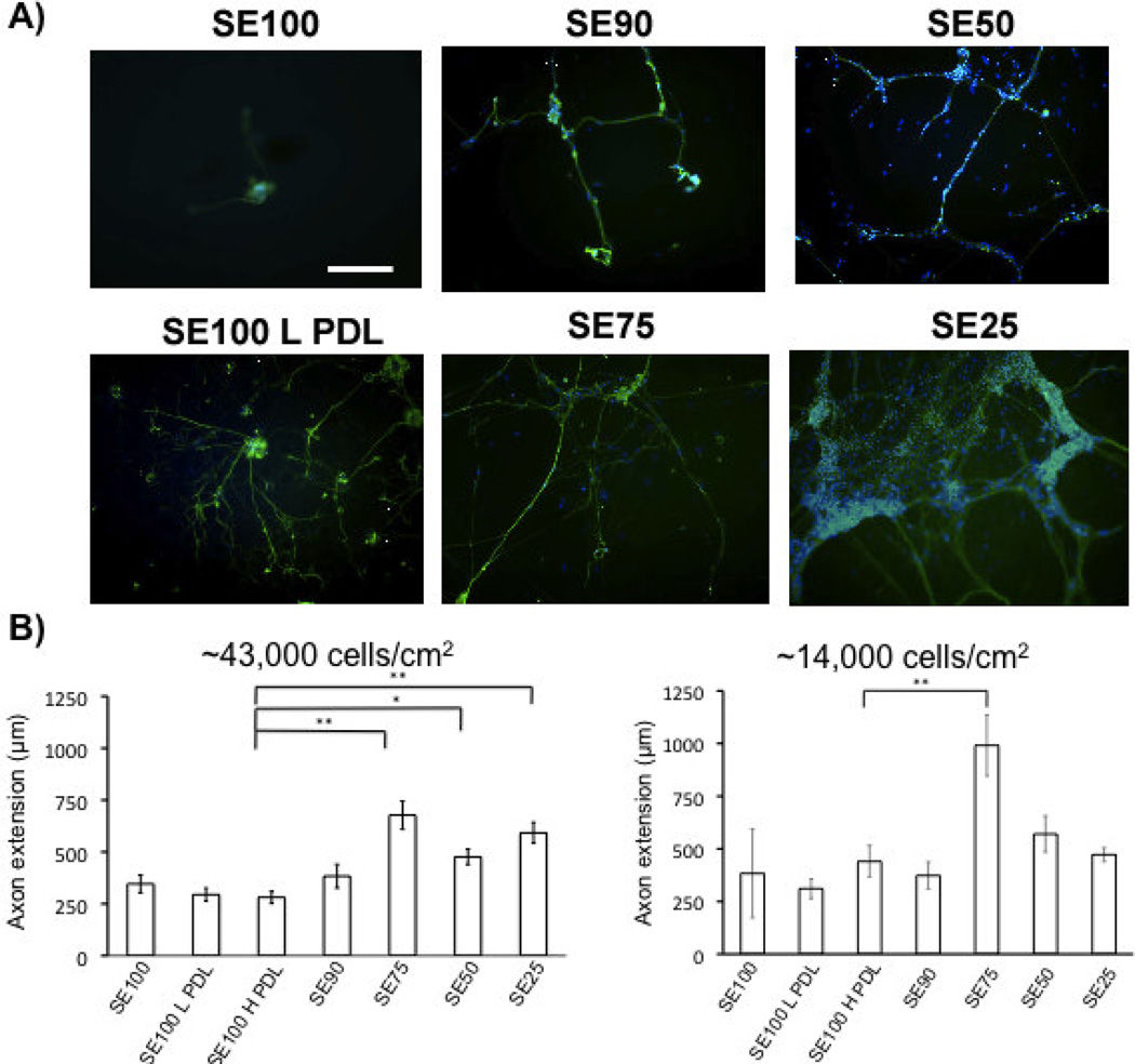
Figure 2.
A) βIII-tubulin (green) staining of DRG neurons grown on silk-tropoelastin films at day 4 (scale bar 250 µm). Nuclear staining with DAPI (blue). B) Quantification of axon extension (For high density, N=30 neurites were measured for each film condition, and taken from six independent fields of view. At low density, isolated neurons were measured, N = 5–10/group). Protein films with tropoelastin contents higher than 25% (w/w) promoted longer axon growth than silk and PDL-coated silk conditions. Significant differences between silk-tropoelastin films and S100 were determined using ANOVA followed by Dunnett’s post hoc test (P*<0.05, P**<0.01).

