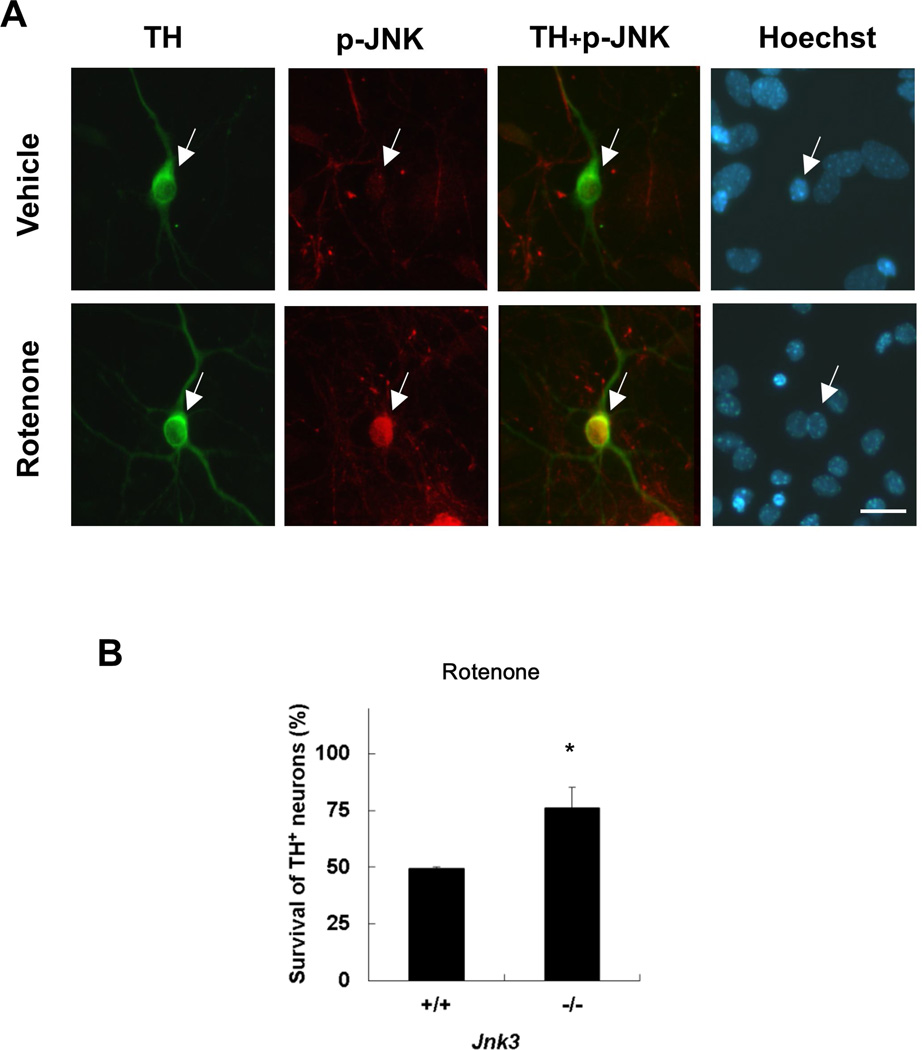
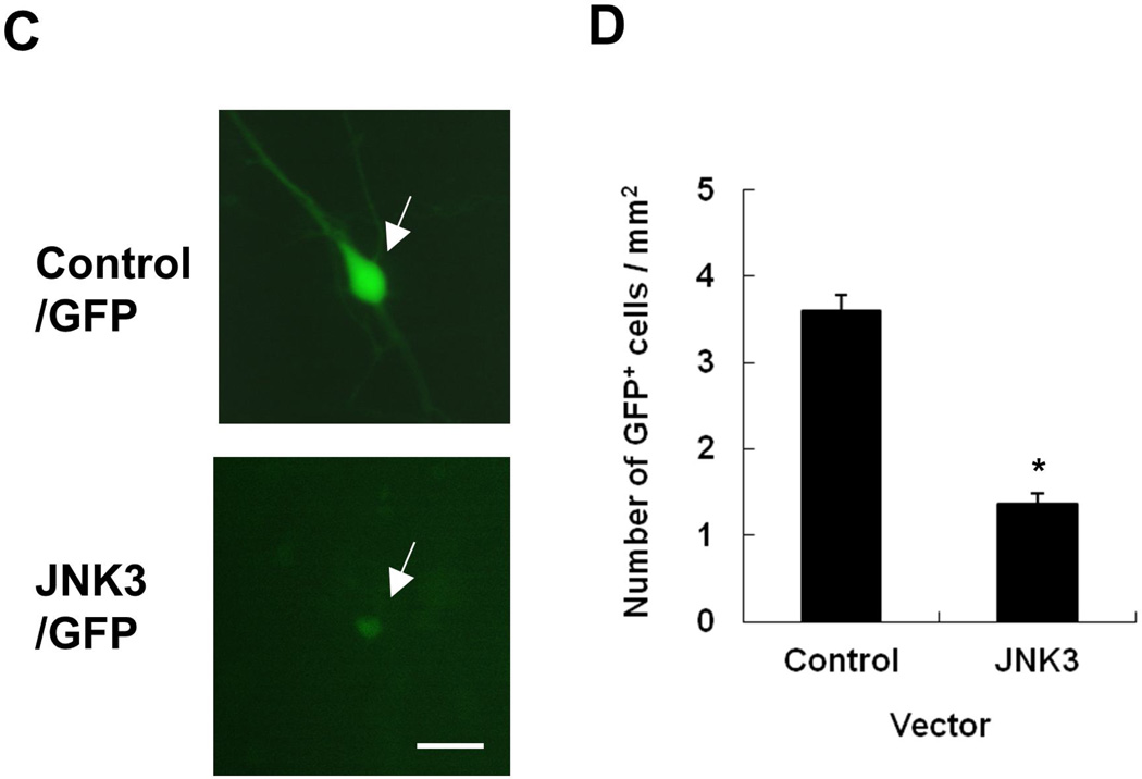
Fig. 1. Rotenone-induced dopamine neuron death requires the function of JNK3, a neural specific JNK isoform.
(A) Primary ventral mesencephalic cultures were prepared from embryonic day (E) 14 wild type C57BL/6 mice. After 6 days in vitro (DIV6), cultures were treated with 5 nM rotenone or vehicle for 8 h. Images are representative photomicrographs immunostained for phosphorylated (p-) JNK and tyrosine hydroxylase (TH) (arrows), a marker for dopamine neurons. Scale bar: 20 µm. (B) TH+ dopaminergic neurons from JNK3−/− mice exhibit resistance to rotenone toxicity. Jnk3+/− mice were mated and primary mesencephalic neurons were cultured separately from each mouse E14 fetus for 6 days. Cells were then treated with 5 nM rotenone for 24 h. (C, D) Ectopic expression of JNK3 induces cell loss. Mesencephalic cultures, prepared from E14 C57BL/6 mouse embryos, were co-transfected with plasmids encoding GFP and JNK3 cDNA or empty vector. Transfected cells were identified by GFP autofluorescence (arrows) 48 h later (C), and the total number of GFP+ cells on each coverslip (9mm diameter) was quantified and presented as cell number / area. (D).


