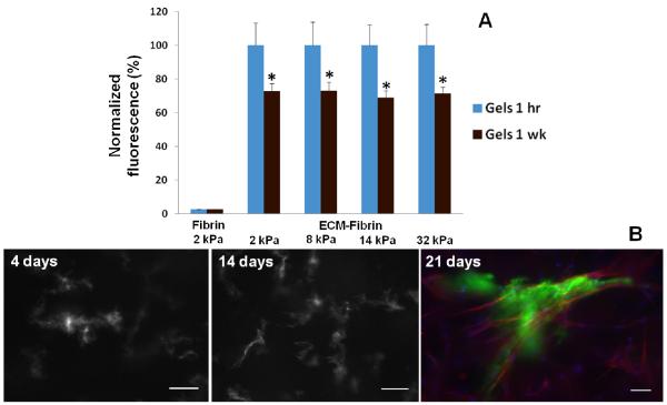
Figure 2. ECM retention within hybrid gels.
(A) Gels containing Alexa Fluor 488 labeled ECM had similar levels 1 hr after gelling. After 1 wk incubation with PBS, fluorescence levels within gels significantly decreased from corresponding baseline levels but were similar in all gels regardless of TG crosslinking. Fluorescence values are normalized to baseline levels at 1 hr (N = 6 per condition). (B) Representative images of AF488 labeled adult ECM-fibrin hybrid gels crosslinked with 1.2 μg/ml TG. ECM becomes more concentrated and fibers become apparent, suggestive of cell interaction and remodeling. Day 21 shows ECM (green), cells labeled with TRITC-phalloidin (red) and Hoechst stain for nuclei (blue). Scale bars = 25 μm.

