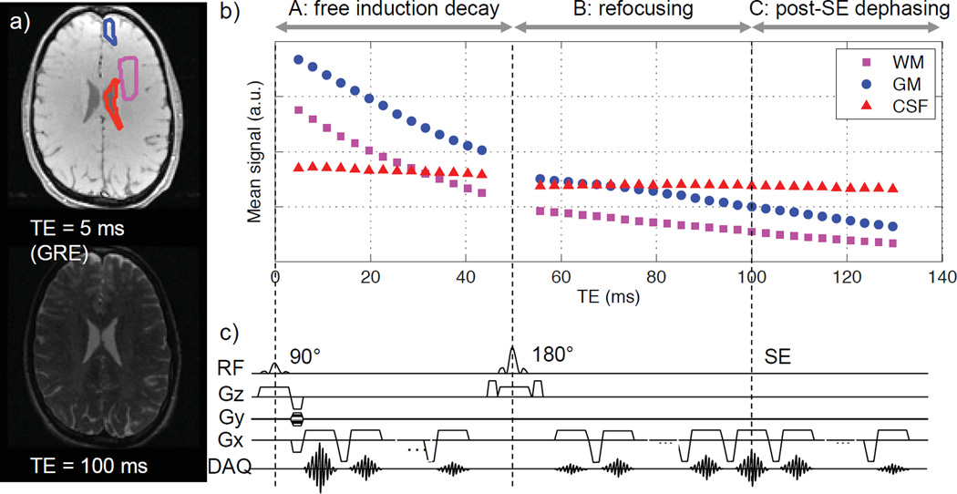
Figure 1.
Modified GESFIDE sequence used in this study: a) representative gradient echo and spin echo images; b) representative signal evolution of ROIs in WM, GM and CSF; c) sequence diagram. The original GESFIDE sequence only samples parts A and B of signal evolution, while this modified version continues readout to sample echoes after the SE (part C), thus acquiring the necessary data points for the GESSE technique.

