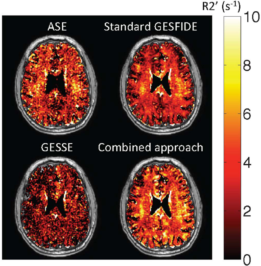
Figure 3.
Representative R2’ maps (superimposed on T1-weighted anatomic images) obtained using standard measurement methods. Visual inspection shows that R2’ values measured by GESSE are lower than those from other techniques. This is supported by quantitative results (Table 2). All methods produced a few voxels in the sulci with small negative R2’ values. These voxels were included in subsequent analysis.

