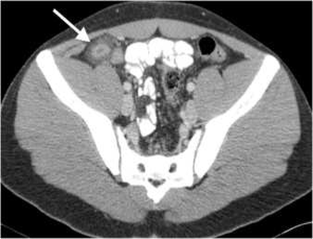
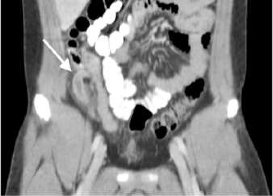
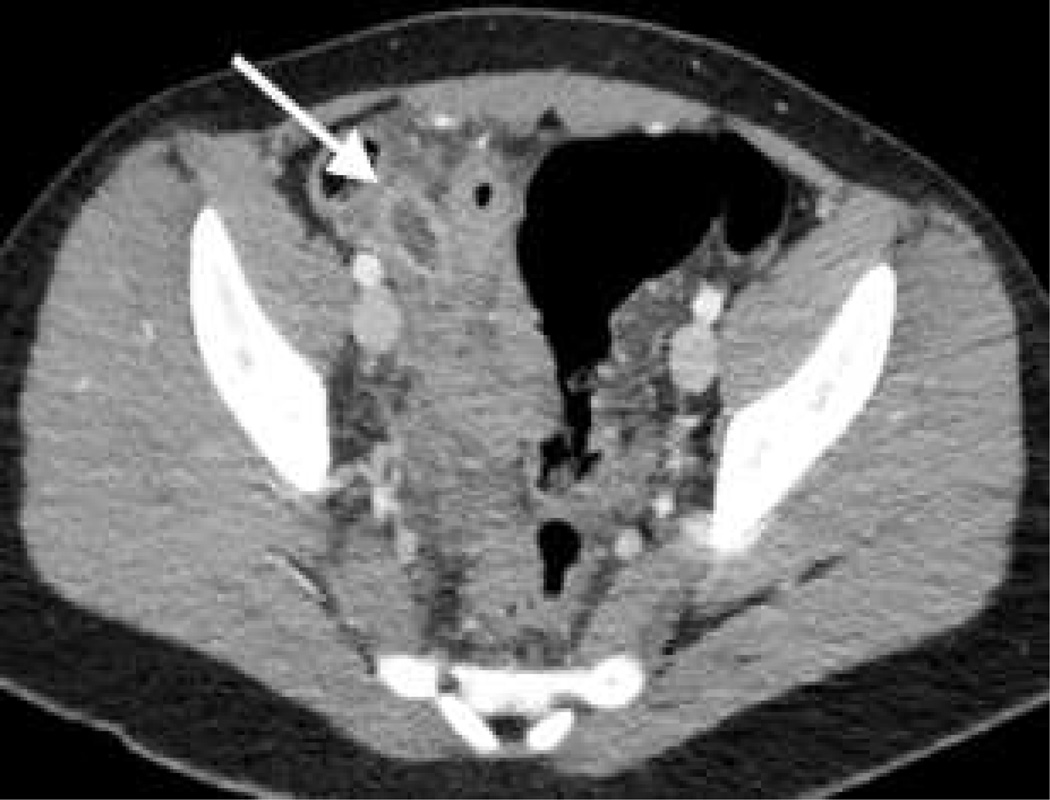
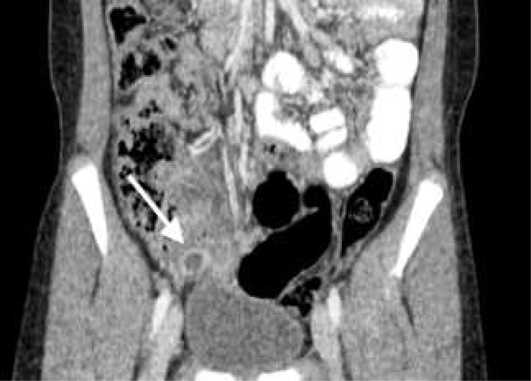
Fig. 1.
Axial (a) and coronal (b) contrast-enhanced CT images of the pelvis in a 15-year-old boy show a 14-mm diameter appendix (arrow) with periappendiceal fat stranding, indicating presence of inflammation (group A, traditional pediatric weight-based protocol with filtered back projection reconstruction; 3 mm axial and coronal section thickness; CTDIvol 16 mGy; SSDE 23 mGy). Axial (c) and coronal (d) contrast-enhanced CT images of the pelvis in a 12-year-old boy show a 14-mm diameter, fluid-filled appendix (arrow) and periappendiceal inflammation (group B, filtered back projection/iterative reconstruction technique blend; 3 mm axial and coronal section thickness; CTDIvol 3 mGy; SSDE 5 mGy). Surgical pathology confirmed appendicitis in both patients


