Figure 1.
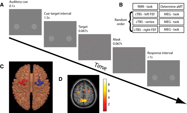
Experimental paradigm and setup. A, Experimental paradigm. Each trial began with an auditory cue (high or low tone) instructing participants to covertly attend to the luminance pedestal in either hemifield. Left and right attentional cues were presented pseudorandomly. Following a 1.5 s interval oriented Gabor patches appeared in both pedestals. Participants had to discriminate the orientation of the cued patch (45° clockwise or anticlockwise from vertical) while ignoring the uncued distracter patches (0° or 90° from vertical). In the fMRI version of this task, used to localize the FEF for TMS, participants completed short (22 s) blocks consisting of spatially cued trials contrasted with blocks of nonspatially cued trials. B, Experimental time course. Participants completed four experimental sessions. In Session 1, left and right FEF were functionally localized based on the attention task performed in the MRI scanner, and the active motor threshold to titrate TMS intensity was determined for each individual. Sessions 2, 3, and 4 consisted of cTBS to one of the three target sites (order counter-balanced across participants and separated by at least 1 week) followed by performance of the attention task during MEG recordings. C, fMRI localizer. Individual TMS target sites for left and right FEF stimulation are shown, superimposed on a standard brain, as derived from the peak voxels within anatomical constraints of the “shift attention” versus “hold attention” contrast of the fMRI localizer task. D, Group fMRI activation map for the contrast SHIFT > NO-SHIFT (t values). Map is thresholded at p < 0.001 (uncorrected).

