
Figure 4. Histological staining of iPSC-derived and native mouse femoral cartilage following treatment with IL-1α.
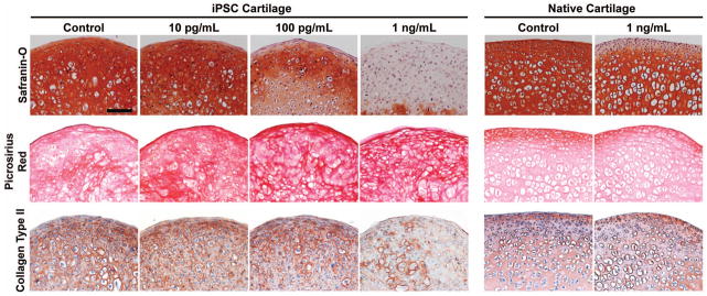
Safranin-O staining of iPSC and native cartilage, indicating a loss of GAG content from both tissues following IL-1α treatment at ≥100 pg/ml. Pircrosirius red staining indicated extensive collagen content in all samples, and no significant changes following cytokine treatment. While there was no change in total collagen content, a loss of collagen II immunostaining was seen, but only in iPSC cartilage treated with 1 ng/ml of IL-1. (Scale bar = 100 μm)