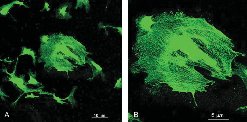
Figure 1.
Fluorescence microscopy of human retinal pericytes (HRP) shows labeling of α-smooth muscle actin fibers. Pericytes were immunoreacted with anti-α- smooth muscle actin conjugated with FITC. Labeled fibers illustrate the identity and phenotype of HRP. (A) Field of labeled HRP. Bar is 10 μm. (B) Close-up of a single HRP clearly showing α-smooth muscle actin fibers. Bar is 5 μm.

