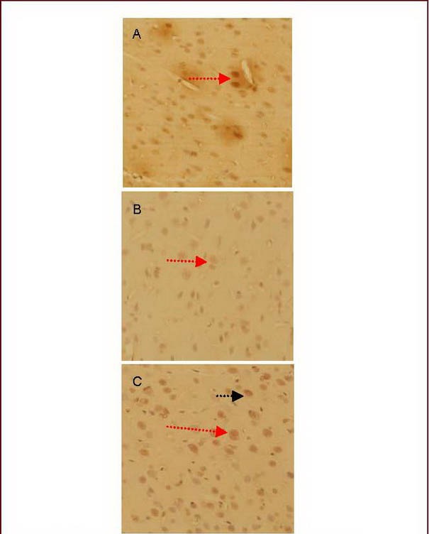
Figure 1.

Morphology of the corticotropin release hormone (CRH)-positive neurons in the hypothalamic paraventricular nucleus (light microscopy, diaminobenzidine staining, ×400).
(A) Model group.
(B) Anti-CRH serum group.
(C) Electroacupuncture + anti-CRH serum group.
The CRH immunoreactive substances exhibited yellow granules in the hypothalamus (red arrow), gathering in the perinuclear cytoplasm.
The CRH-positive neurons exhibited spindle and spherical shapes. Some positive cell bodies had one or two dendrites (black arrow). The cell body and dendrites were stained yellow.
