
Figure 5.
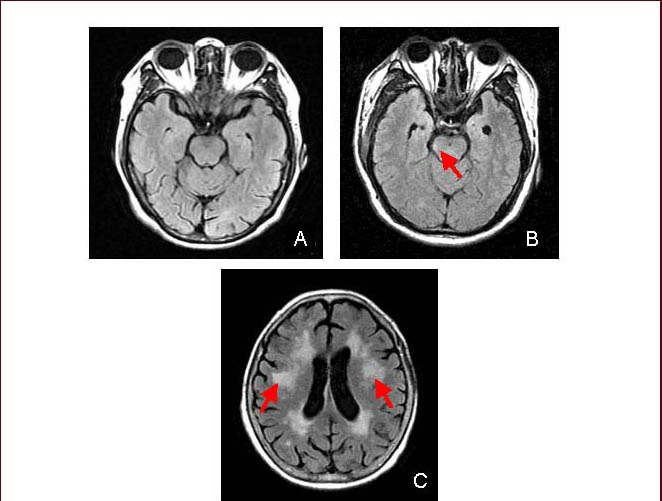
Fluid-attenuated inversion-recovery (FLAIR) images from healthy, axonal injury and demyelinating disease subjects.
(A) FLAIR image of brain from a healthy subject (male/23 years old).
(B) Image from an axon injury patient (male/25 years old), focused on abnormally high signal in the midbrain area, shown as red arrow points.
(C) Image from a 65-year-old man with demyelinating disease. Red arrows represent lesions.