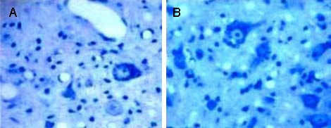
Figure 4.

Nissl bodies in the injured spinal cord of rats in each group (Nissl staining, light microscope, × 400).
Nissl bodies in the model group (A) were significantly reduced, compared with the experimental group (B).

Nissl bodies in the injured spinal cord of rats in each group (Nissl staining, light microscope, × 400).
Nissl bodies in the model group (A) were significantly reduced, compared with the experimental group (B).