
Figure 2.
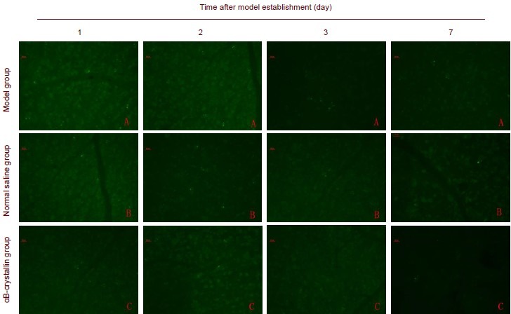
Caspase-3 expression in retinal ganglial cells (RGCs) in rats (immunofluorescence staining, × 200).
Following acute ocular hypertension, Caspase-3 positive expression is detected in the rat retina, mainly in the cytoplasm, and gradually decreased by 2 days. Green fluorescence spots represents caspase-3-positive cells. Fluorescein served as the fluorescent dye.