
Figure 1.
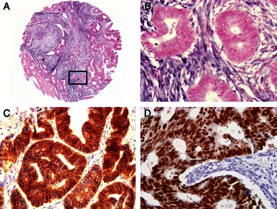
In situ hybridization for miR-21 and immunohistochemistry for E-cadherin and MTA1. (A) A representative 2 mm tumor tissue core from the colorectal cancer tissue microarray shows diffuse strong miR-21 expression in the stroma. (B) High-magnification image of insert in (A) shows that miR-21 signals are strong in the stromal cells of colorectal cancer but not in the tumor cells. Magnification x400. (C) Tumor cells show strong membranous expression of E-cadherin. Magnification x400. (D) Tumor cells show strong nuclear expression of MTA1. Magnification x400.