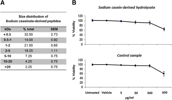
Figure 1.

Peptide size distribution of the sodium casein-derived peptides and effects on endothelial cell viability . (A) Gel permeation chromatography of the sodium casein-derived peptides was performed to assess the peptide size distribution of the hydrolysate. Data show the mean ± SEM of four incubation reactions. (B) MTT assay was carried out to determine the cell viability of EC treated with casein hydrolysate or with the control sample for 18 h, resulting in more than 94% ±3.4% viable cells up to a concentration of 300 μg/ml. Data are expressed as mean ± SEM of 3 independent experiments. Data were reported as percentage of control (untreated cells).
