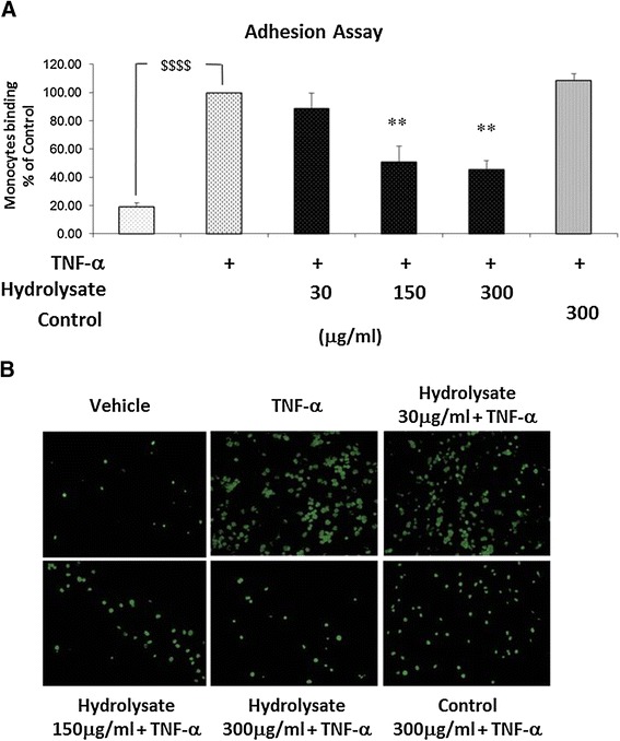
Figure 3.

Adhesion of human monocytes to TNF-α activated EC is prevented by sodium casein-derived peptides. EC were treated with samples for 18 h, followed by 6 h stimulation with TNF-α 0.5 ng/ml) and a static adhesion assay with fluorescence-labelled THP-1 human monocytes was performed. (A) Adherent monocytes were measured in a plate fluorescence reader with 485 nm excitation and 530 nm emission wavelength. Data were calculated as mean +/− SEM of 3 independent experiments. Statistical analysis was carried out using one-way ANOVA employing Dunnett correction for multiple comparisons. A statistical value of *P < 0.05 or greater was considered significant; $$$$ (p < 0.0001) vehicle vs control; **P < 0.01 treatments vs control (TNF-α activated EC). (B) Representative fluorescence microscopy photomicrographs of monocytes adhesion to EC are shown: vehicle (top left); TNF-α (top centre); hydrolysate 30 μg/m l+ TNF-α (top right); hydrolysate 150 μg/ml + TNF-α (bottom left); hydrolysate 300 μg/ml + TNF-α (bottom centre); control 300 μg/ml + TNF-α (bottom right).
