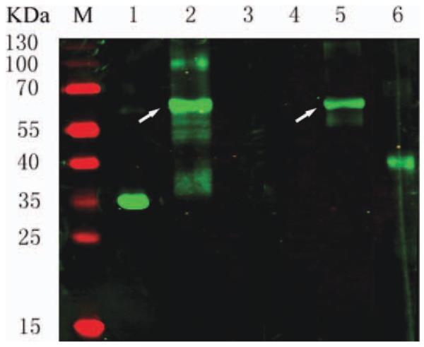
FIGURE 3.
Western blot analysis of fusion protein. Lanes 1–3 were detected with an anti–c-myc tag MAb9E10; Lanes 4–6 were probed with an anti-DAF MAb. Controls included scFv1956 (lanes 1 and 4) and DAF (lanes 3 and 6). The white arrows on lanes 2 and 5 indicate the fusion protein scFv-DAF. [Color figure can be viewed in the online issue, which is available at wileyonlinelibrary.com.]

