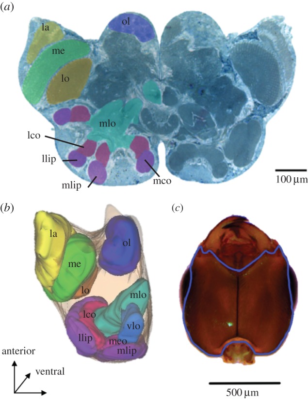
Figure 1.

Acacia ant (Pseudomyrmex spinicola) transverse brain section and ventral view of head. (a) Brain section highlighting some of the measured neuropiles including: the lamina (la), lobula (lo) and medulla (me) of the optic lobe and the olfactory lobe (ol). In the mushroom bodies, we measured the lip and collar of the lateral (llip and lco) and medial calyces (mlip and mco); the vertical lobe (alpha; not visible) and medial lobe (mlo, beta). (b) Neuropile dimensions of brain sections were used to generate three-dimensional reconstructions of the brain regions to obtain volumetric measurements. Colours correspond to the neuropiles shown in the section; the vertical lobe (vlo), not visible in the two-dimensional section, is shown here. (c) Ventral view of the head showing in blue the contour used to calculate the head area, excluding eyes and mouthparts. (Online version in colour.)
