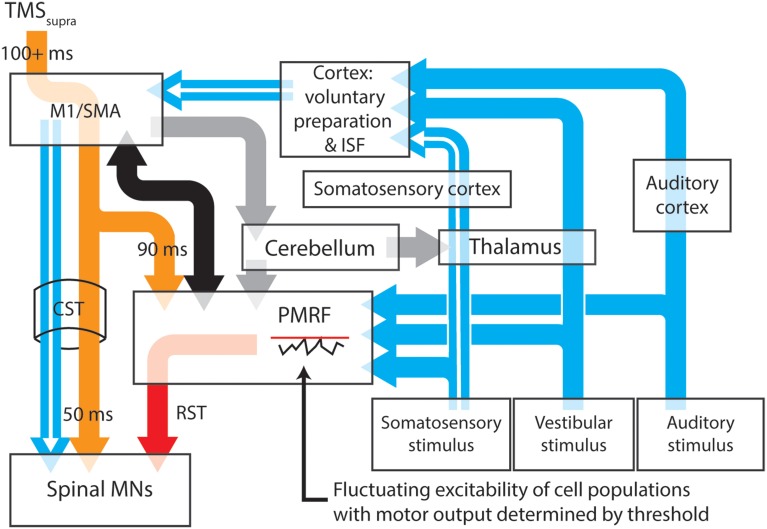
Figure 1.
A proposed model for the neural pathways subserving the expression of long latency stretch reflexes, startle and startReact responses. Transcortical contributions to long latency stretch reflexes are enabled by a pathway that involves the primary somatosensory and motor cortices before descending to muscles through the corticospinal tract (white arrows). Stimulation of somatosensory, vestibular or auditory systems results in transmission of these signals to the cortex and the pontomedullary reticular formation (blue arrows), each of which have thresholds for activating output cells. Inhibition of the primary motor cortex (or areas involved in movement preparation) by application of suprathreshold TMS (orange arrows) results in almost simultaneous inhibition of cells within the spinal cord and PMRF, although for different periods of time. Spinal motoneurons appear to be inhibitied for ~50 ms after TMS, while cells within the PMRF are inhibited for ~90 ms in non-human primates. Output cells within the motor cortex however, can be inhibited for up to 200 ms (Strick, 1983). During periods of cortical inhibition, activation of the corticospinal tract is not possible, although a combination of cortico-reticulospinal input (black arrows) and sufficiently large sensory input can still activate reticulospinal tract cells (red arrow) after any TMS-induced inhibition of PMRF ceases. The likelihood of PMRF output (startle or startReact responses) is determined in this model by the magnitude of the sensory input and the instantaneous excitability of PMRF cells. A similar situation is likely to exist at the cortical level (see Alibiglou and MacKinnon, 2012). Task, posture and stability-dependent regulation of stretch reflex and startReact responses likely involves input from the cerebellum to both the primary motor cortex and reticular formation (gray arrows). In this figure thalamocortical projections to regions responsible for voluntary motor preparation, but projections directly to M1 may also play an important role in modulating rapid responses. Output nuclei and detailed information about synapse locations for each pathway have been omitted from this figure. M1, Primary motor cortex; SMA, supplementary motor area; PMRF, pontomedullary reticular formation; ISF, intersensory facilitation; MN, motoneuron.

