Figure 1.
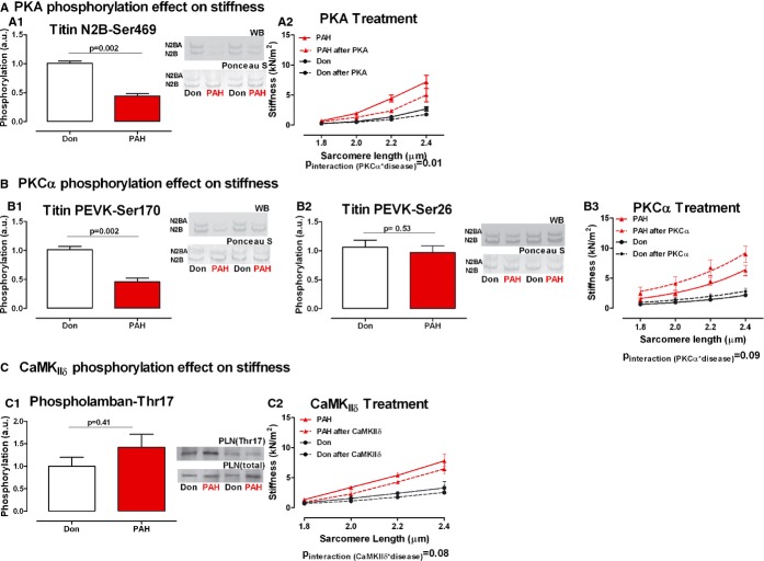
PKA, PKCα, and CaMKIIδ treatment effect on diastolic stiffness mediated by titin phosphorylation. A‐A1. Titin N2B domain serine 469 PKΑ‐dependent phosphorylation. Typical example of the 2 titin isoforms (upper band: N2BA; lower band N2B) immunostained with phosphospecific antibody against serine 469 site on titin N2B domain and the corresponding Ponceau S staining for total titin (nDon=7, nPAH=5). A2. Donor and PAH cardiomyocyte stiffness was measured in relaxing solution at increasing sarcomere length (1.8 to 2.4 μm)—continuous line. The same cardiomyocyte was further incubated with PKA active subunit and stiffness measurements were repeated—dotted line (nDon=4, nPAH=4). B‐B1. Titin PEVK domain serine 170 PKCα‐dependent phosphorylation. Typical example of the 2 titin isoforms (upper band: N2BA; lower band N2B) immunostained with phosphospecific antibody against serine 170 site on titin PEVK domain and the corresponding Ponceau S staining for total titin (nDon=7, nPAH=5). B2.Titin PEVK domain serine 26 PKCα‐dependent phosphorylation. Typical example of the 2 titin isoforms (upper band: N2BA; lower band N2B) immunostained with phosphospecific antibody against serine 26 site on titin PEVK domain and the corresponding Ponceau S staining for total titin (nDon=7, nPAH=5). B3. Donor and PAH cardiomyocyte stiffness was measured in relaxing solution at increasing sarcomere length (1.8 to 2.4 μm)—continuous line. The same cardiomyocyte was further incubated with PKCα active subunit and stiffness measurements were repeated—dotted line (nDon=3, nPAH=3). C‐C1. Phospholamban threonine 17 CamKIIδ‐dependent phosphorylation was used as an indirect measurement of titin CamKIIδ phosphorylation. The phosphorylation level was normalized to the total amount of Phospholamban present in the sample (nDon=7, nPAH=11). C2. Donor and PAH cardiomyocyte stiffness was measured in relaxing solution at increasing sarcomere length (1.8 to 2.4 μm)—continuous line. The same cardiomyocyte was further incubated with CamKIIδ and stiffness measurements were repeated—dotted line (nDon=3, nPAH=3). Data presented as mean±SEM. CamKIIδ indicates calmoduling‐dependent‐kinase; PAH, pulmonary arterial hypertension; PKA, protein‐kinase‐A; PLN, phospholamban.

