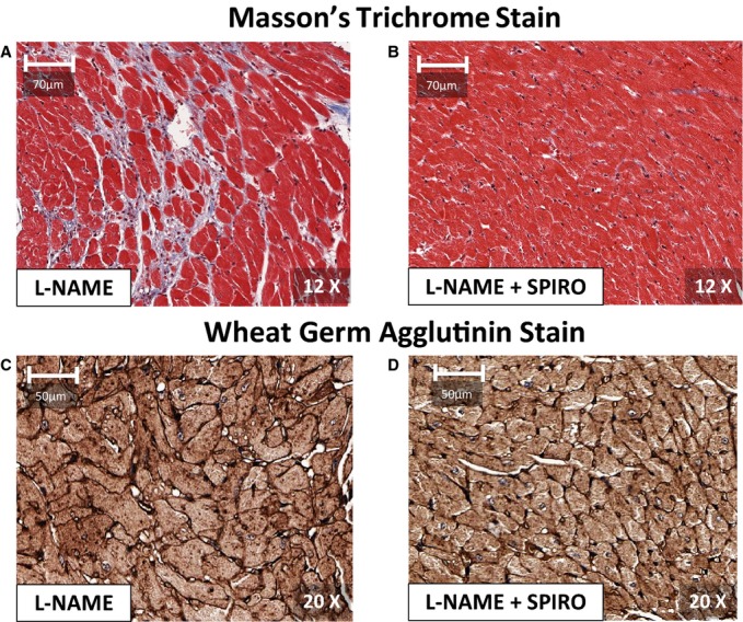
Figure 4.

Short‐axis, mid‐level sections of cardiac tissue from the left ventricle (LV) in mice treated with l‐NAME (A and C), and spironolactone and l‐NAME (B and D), after 7 weeks of l‐NAME. Short‐axis sections stained with Masson's trichrome stains in (A) and (B) illustrate relative abundant interstitial fibrosis (appearing blue) in l‐NAME, as compared to l‐NAME+spironolactone. Adjacent slices in the same mice in (C) and (D) were stained with fluorescein isothiocyanate‐conjugated (FITC) wheat germ agglutinin to delineate cell membranes. With l‐NAME alone, mice showed cardiomyocyte hypertrophy, compared to l‐NAME+spironolactone. The cardiomyocyte diameters in the anterior, lateral, inferior and septal LV segments were significantly larger in l‐NAME compared to l‐NAME+spironolactone (P<0.001). l‐NAME indicates Nω–nitro‐l‐arginine‐methyl‐ester.
