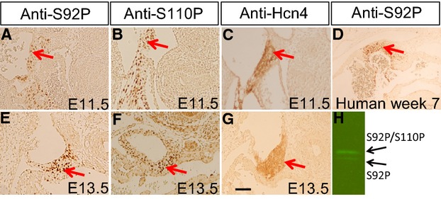
Figure 3.

Presence of p‐Shox2a in the SAN of mouse and human embryos. Immunohistochemical staining using anti‐S92P (A, E) or anti‐S110P (B, F) specific antibodies demonstrates the presence of p‐Shox2a in the SAN of E11.5 (A, B) and E13.5 (E, F) mouse embryo. Immunohistochemical staining shows the expression of Hcn4 in the SAN of E11.5 (C) and E13.5 (G) mouse embryo. D, Immunohistochemical staining using anti‐S92P antibody reveals the presence of p‐SHOX2a in the SAN of human embryo at 7‐week gestation. F, Western blotting using anti‐S92P antibody reveals the presence of p‐Shox2a in E11.5 mouse embryonic hearts. Red arrows point to the SAN region. Scale bar=20 μm for all panels. SAN indicates sinoatrial node.
