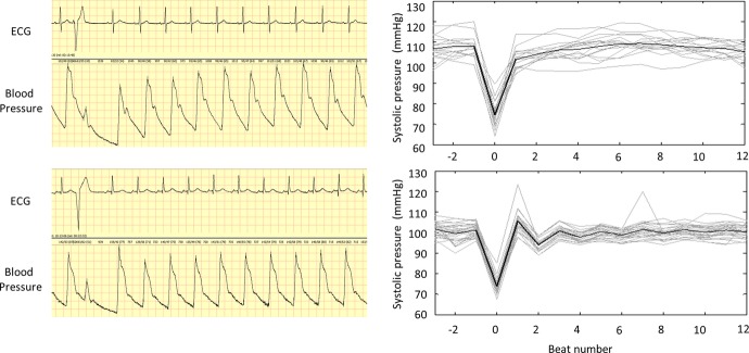
Figure 1.

Determination of PESP based on ECG and BP signals. Left panel shows ECG and blood pressure signals of a patient surviving the follow‐up period (upper tracing) and a patient who died 16 months after the index MI (lower tracing). The systolic pressure of the first postectopic beat in the upper tracing is clearly lower than that of the following 9 beats, indicating that PESP is not present. Conversely, the systolic pressure of the corresponding beat in the lower tracing is noticeably higher than that of the subsequent beats (PESP is present). Right panels show peak systolic pressure of consecutive heart beats before, during, and after ectopic beats in these patients. Individual episodes are shown in gray, their average in black. MI indicates myocardial infarction; PESP, postextrasystolic potentiation.
