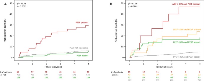
Figure 3.

Kaplan‐Meier's curves for the association of PESP and mortality. A, Mortality probability over 5 years in patients stratified according to the presence (red curve) and absence of PESP (green curve). The mortality probability of patients whose recordings lacked VPCs suitable for the quantification of PESP is also shown (gray curve). Because there was no significant difference in patients with PESP absent and without suitable VPCs, both subgroups were merged for additional analyses. B, Probability of death in patients stratified by the combination of PESP (absent and present) and LVEF (≤35% and >35%). The numbers of patients at risk in the individual groups at 0, 1, 2, 3, 4, and 5 years are shown below the graphs in the same color coding. P values for the overall comparison at 5 years are indicated. LVEF indicates left ventricular ejection fraction; PESP, postextrasystolic potentiation; VPC, ventricular premature complex.
