Figure 6.

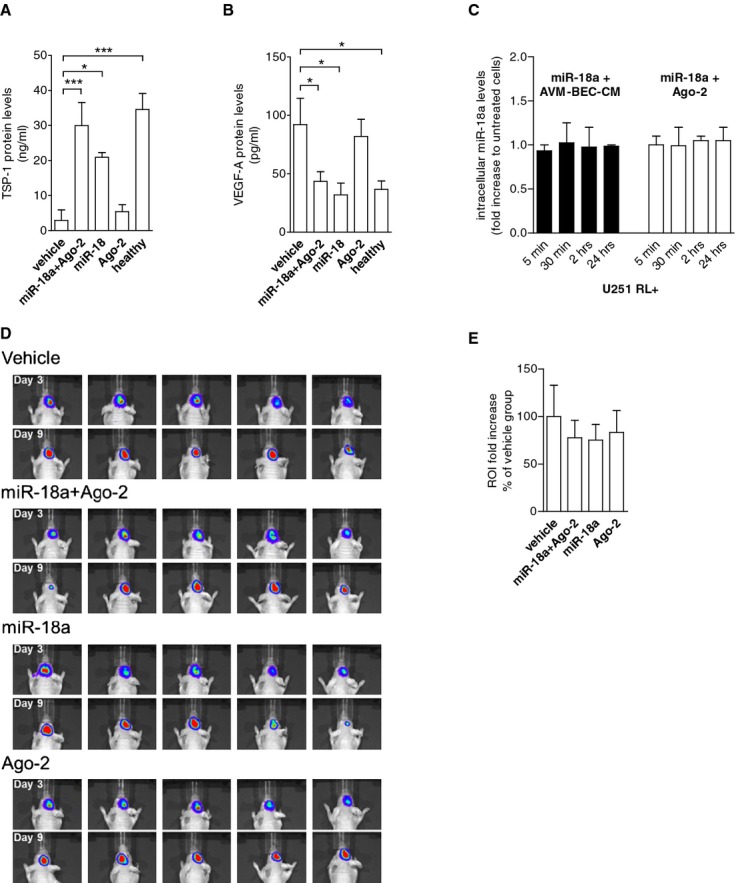
Co‐treatment of miR‐18a and Ago‐2 in vivo “normalizes” TSP‐1 and VEGF‐A plasma levels. A, Athymic nude mice were implanted with glioma cells intracranially. After 3 days, animals were treated intravenously with vehicle, miR‐18a plus Ago‐2, miR‐18a alone or Ago‐2 alone every 48 hours for 3 cycles. Subsequently, plasma was tested for TSP‐1 (A) and VEGF‐A (B). miR‐18a and Ago‐2 combination treatment caused the most significant increase of TSP‐1 levels (n=5; *P<0.05; ***P<0.001; paired 1‐way ANOVA followed by Dunnett's Multiple Comparison Test), and reduction of VEGF‐A levels (n=5; *P<0.05; paired 1‐way ANOVA followed by Dunnett's Multiple Comparison Test). Control plasma (healthy) was obtained from normal athymic mice. C, qPCR analysis of miR‐18a detection showed that tumor cells did not internalize miR‐18a in the presence of AVM‐BEC‐CM (black bars) or Ago‐2 (white bars) (n=3). D, Imaging of intracranial renilla luciferase‐positive tumor cells day 3 after implantation (beginning of treatment) and day 9 (treatment completion). E, Region of interest (ROI) analysis of detected luminescence showed that miR‐18a and Ago‐2 combination and miR‐18a treatment alone led to slower tumor growth as compared to vehicle‐treated animals, albeit not statistically significant (n=5). Data are presented as mean±standard error of the mean (SEM). ANOVA indicates analysis of variance; AVM, arteriovenous malformation; Ago‐2, Argonaute‐2; BEC, brain endothelial cells; CM, conditioned media; qPCR, quantitative real‐time polymerase chain reaction; TSP‐1, thrombospondin‐1; VEGF, vascular endothelial growth factor.
