
Figure 3.
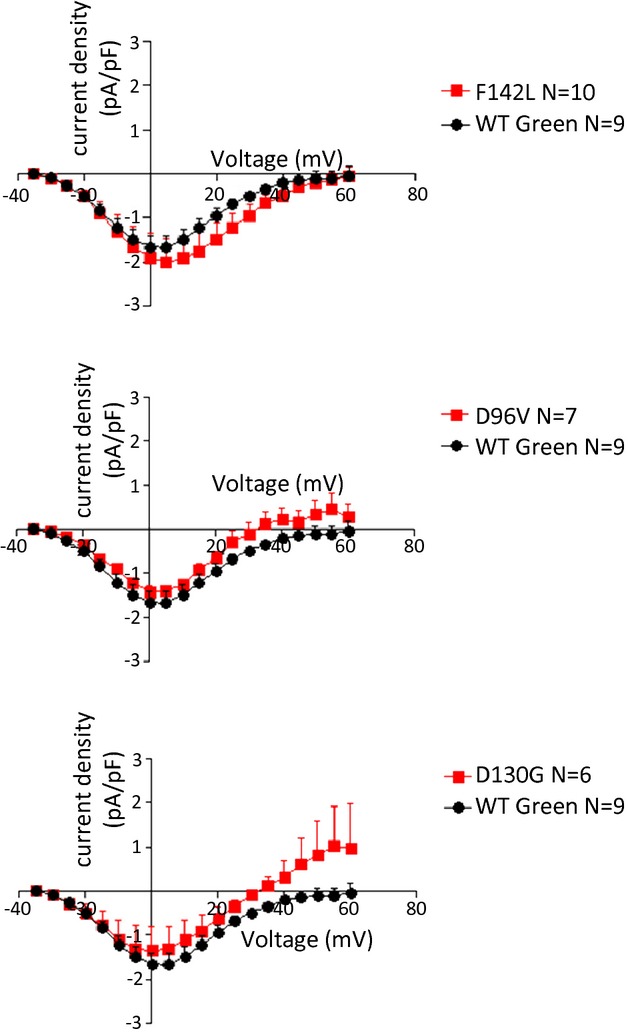
Peak current‐voltage relationships for ICa,L in fetal ventricular cardiomyocytes. WT calmodulin data are repeated in each panel for clarity of presentation. No significant differences were noted in voltage dependence or amplitude. See Table 3 for Boltzmann distribution fitted parameters. WT indicates wild type.